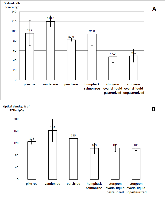
Figure 2. The effect of the lyophilized Siberian sturgeon ovarian liquid and the extracts of various fish species upon the senescence (A) (the percentage of the SA-β-Gal-positive cells) and the proliferation (B) of the LECH-T cells. A: for the SIPS induction, the cells were treated with 50 μM Н2О2 for 1 h and later stained for β-Gal. The result is expressed as the ratio of the proportion of stained cells after incubation with the sample to the proportion of stained cells treated with Н2О2 without further addition of the sample. B: cell proliferation was assessed at 96 h after SIPS induction using crystal violet. The result is expressed as the ratio of the optical density after incubation with the sample to the optical density of cells treated with Н2О2 without further addition of the sample (mean ± SD). The difference was considered as statistically significant of р < 0.05