|
|
Identification of Genes whose mRNAS are Subjected to Alternative Splicing
by Endonuclease EndoG Action in Human and Murine CD4+ T Lymphocytes
D.D. Zhdanov1,2*, N.S. Novachly2, M.V. Pokrovskaya1, S.S. Aleksandrova1, T.A. Kabardokov2,
N.N. Sokolov1
Key words: lymphocytes; EndoG; alternative splicing; sequencing
Alternative splicing (AS) of mRNA is one of the ways of regulation of protein functions (including enzymatic activity) [1]. To date many of proteins which regulate this process have been identify; however precise mechanism of AS remains to be investigated. Previously, we showed that apoptotic endonuclease EndoG was capable of inducing AS of mRNAs of telomerase catalytic subunit hTERT (human Telomerase Reverse Transcriptase) [2, 3] and deoxyribonuclease 1 (DNase I) [4] in human lymphocytes. In both cases induction of AS led to inhibition of the enzymatic activity. Later, the ability of EndoG to induce AS have been shown using mice and rat lymphocytes [5]. Thus, EndoG can be considered as a potent modulator of AS. Its mechanism of action is still poorly understood. It is of interest to study the ability of EndoG to modulate AS of mRNA of other genes. In this paper we have identified human and murine genes whose mRNAs are alternatively spliced by EndoG.
Venous blood samples of four healthy donors were collected into tubes with K3EDTA as an anticoagulant («Greiner Bio-One», Kremsmünster, Austria). Blood samples from C57BL/6 mice and Wistar rats («Pushchino Breeding Center for Laboratory Animals», Pushchino, Russia) were obtained by cardiac puncture after euthanasia with CO2. CD4+ T cell purification was performed by magnetic selection using the human CD4+ Isolation Kit («Miltenyi Biotec», Germany) according to the manufacturer’s protocol. CD4+ cells were seeded at a concentration of 5 × 10⁵ cells/mL of the RPMI-1640 culture medium («Life Technologies», USA) containing 10% FBS (Fetal Bovine Serum, «Thermo Fisher Scientific Inc.», Waltham, USA), growth factors IL-2 (100 U/mL, «R&D Systems», USA), anti-CD3 antibodies (5 μg/mL, «MedBioSpectrum», Russia), and anti-CD28 antibodies (2 μg/mL, «eBioscience Inc.», USA) [6]. Isolation of CD4+ T cells from murine and rat blood was performed using the CD4+ T Cell Isolation Kit, mouse («Miltenyi Biotec», Germany). CD4+ T cells from mice and rats were cultured in the above described medium with addition of 1 mM sodium pyruvate («Applichem», Germany), 10 mM HEPES («Sigma-Aldrich», USA), and 0.02 mM 2-mercaptoethanol («Sigma-Aldrich», USA). Cells were cultivated in a CO2 incubator at 37°C, 5% CO2 and 90% humidity. Transfection of human, murine and rat CD4+ T cells was performed by one of the following plasmids: Human pEndoG-GFP plasmids, Mouse pEndoG-GFP, Rat pEndoG-GFP, respectively, or the control plasmid pGFP. Plasmids were prepared using the vector pGFP-N1 («Clontech», USA). Transfection was performed using Lipofectamine 2000 («Invitrogen», USA) according to the manufacturer’s protocol. Transfection efficiency was estimated by flow cytometry by counting GFP-positive cells labeled with CD4-VioBlue («Miltenyi Biotec», Germany) using cytometr MACS Quant Analyzer 10 («Miltenyi Biotec», Germany). To induce DNA damage and EndoG expression, CD4+ T cells were cultivated with 60 μM cisplatin (cis-diamine dichloroplatinum (II), «Sigma-Aldrich», USA) for 48 h [3, 7]. Determination of number of cells with damaged DNA was performed by the TUNEL assay (Terminal deoxynucleotidyl transferase-mediated d-UTP Nick End Labeling) [8] using the FlowTACSTM Apoptosis Detection Kit («R&D Systems», USA) for flow cytometry.
Isolation of total RNA and EndoG expression analysis was performed according to the previously described protocol [2].
Total RNA was isolated from transfected, incubated with cisplatin or control cells followed by formation of cDNA libraries performed using Illumina TruSeq Stranded Total RNA Library Prep Kit with Ribo-Zero Human/Mouse/Rat («Illumina Inc.», USA) [9, 10]. Each cDNA sample was diluted to a concentration of 6 pM and sequenced with HiScanSQ («Illumina Inc.», USA) in the Total RNA-Seq application as described in [11]. The sequencing results were analyzed for the UCSC Homo sapiens reference genome hg19, Mus musculus reference genome mm10 and Rattus norvegicus reference genome rn10 using TopHat v1.3.0 [12].
Statistical analysis of the results was performed using Student’s criterion by means of Statistica 9.0 («StatSoft Inc.», USA) software. Results are presented as mean value ± standard error of mean. Numbers were considered statistically significant when p ≤ 0.05.
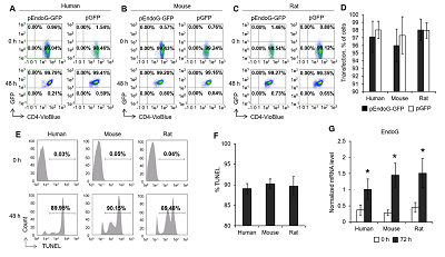
For identification of genes, whose mRNAs are subjected to alternative splicing regulated by EndoG, we have induced EndoG expression by transfection of human, murine and rat CD4+ T lymphocytes with plasmid constructs based on the pGFP-N1 vector. PGFP empty plasmid, which encodes only green fluorescent protein (GFP) was used for control transfection. Induction of EndoG was also performed by incubation of cells with the DNA-damaging agent cisplatin. Transfection efficiency was close to 100% in 48 h after transfection in human, murine and rat lymphocytes (Fig. 1 A–D). The number of cells, having DNA damages, which were determined by the TUNEL assay for flow cytometry, was 88.2–90.5% (fig. 1 E-F). 48 h after incubation with cisplatin, EndoG mRNA was evaluated by real-time RT-PCR. Induction of DNA damage by cisplatin resulted in a significant increase in EndoG expression in lymphocytes of all species studied (Fig. 1G).

|
Figure 1.
Efficiency of transfection and EndoG induction in human, mice and rat lymphocytes. Results of flow cytometry for CD4+ T lymphocytes from human (A), mice (B) and rats (C) lymphocytes 48 h after transfection. Transfection efficiency determined by flow cytometry (D). Number of CD4+ T lymphocytes with damaged DNA, measured by TUNEL for flow cytometry (E). Induction of TUNEL-positive cells 48 h after incubation with cisplatin (F). EndoG mRNA levels in cells determined by real time RT-PCR. mRNAs levels are normalized relative to the expression of the reference 18S gene.
(n = 4).
|
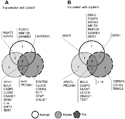
Total cell RNA was sequenced. 30 million of 75-bp paired-end reads were sequenced. Analysis of mRNA levels of splice variants of different genes showed that in human, mouse, and rat cells transfected with pEndoG-GFP, and in cells incubated with cisplatin, the number of mRNA splice variants of 28 genes changed (Fig. 2, Table 1).
|
Figure 2.
Identified genes whose mRNA splice variants levels changed after EndoG overexpression. Comparison of identified mRNA in CD4+ T lymphocytes after transfection with EndoG gene (A) or incubation with cisplatin (B).
|
|
CLOSE

|
Table 1.
Identified genes whose mRNA AS changes upon overexpression of EndoG.
|
Defined shift in the proportion of splice-variants of mRNA was not associated with the changes of total mRNAs of these genes. Transfection of cells with control plasmid pGFP did not lead to the changes in the proportion of mRNA splice-variants of defined genes. Changes in the proportion of mRNA splice variants was confirmed by real-time RT-PCR. Among defined mRNAs in all human, mice and rat lymphocytes, the levels of 9 of them (ARIH1, BCLX, CASP2, CLDN7, DNASE1, GRIN1, IL18, SIRT1 and TERT) changed after transfection with EndoG gene (Fig. 2 A) and 6 (BCLX, CASP2, CLDN7, CLOCK, DNASE1, TERT) after incubation with cisplatin (Fig. 2 B). These results indicate the involvement of EndoG into the function of multiple proteins, which regulate diverse cellular processes.
Thus, we have shown that EndoG participates in modulation of alternative splicing of mRNAs of defined genes in human, mice and rats.
The study was approved by the Ethics Committee of the Institute of Biomedical Chemistry. Written consent was obtained from all donors participating in the research.
The work was performed in the framework of the Program for Basic Research of State Academies of Sciences for 2013-2020.
The authors declare that they have no conflict of interest.
- Kim, E., Magen, A., Ast, G. (2007) Different levels of alternative splicing among eukaryotes. Nucleic Acids Res., 35, 125. DOI
- Zhdanov, D.D., Vasina, D.A., Grachev, V.A., Orlova, E. V., Orlova, V.S., Pokrovskaya, M. V., Alexandrova, S.S., Sokolov, N.N (2017) Alternative splicing of telomerase catalytic subunit hTERT generated by apoptotic endonuclease EndoG induces human CD4 + T cell death. Eur. J. Cell Biol., 96, 653–664. DOI
- Zhdanov, D.D., Vasina, D.A., Orlova, E. V., Orlova, V.S., Pokrovskaya, M. V., Aleksandrova, S.S., Sokolov, N.N. (2017) Apoptotic endonuclease EndoG regulates alternative splicing of human telomerase catalytic subunit hTERT. Biochemistry (Moscow), Suppl. Ser. B Biomed. Chem., 11, 154–165. DOI
- Zhdanov, D.D., Gladilina, Y.A., Pokrovsky, V.S., Grishin, D. V., Grachev, V.A., Orlova, V.S., Pokrovskaya, M. V., Alexandrova, S.S., Plyasova, A.A., Sokolov, N.N. (2019) Endonuclease G modulates the alternative splicing of deoxyribonuclease 1 mRNA in human CD4+ T lymphocytes and prevents the progression of apoptosis. Biochimie, 157, 158–176. DOI
- Zhdanov, D.D., Gladilina, Y.A., Pokrovskaya, M. V., Aleksandrova, S.S., Grishin, D. V., Podobed, O. V., Sokolov, N.N. (2018) Induction of Alternative Splicing and Inhibition of Activity of Telomerase Catalytic Subunit by Apoptotic Endonuclease EndoG in Human T, B, and NK Cells. Bull. Exp. Biol. Med., 164, 478–482. DOI
- Vasina, D.A., Zhdanov, D.D., Orlova, E. V., Orlova, V.S., Pokrovskaya, M. V., Aleksandrova, S.S., Sokolov, N.N. (2017) Apoptotic endonuclease EndoG inhibits telomerase activity and induces malignant transformation of human CD4+ T cells. Biochemistry (Moscow), 82, 24–37. DOI
- Basnakian, A.G., Apostolov, E.O., Yin, X., Napirei, M., Mannherz, H.G., Shah, S. V. (2005) Cisplatin Nephrotoxicity Is Mediated by Deoxyribonuclease I. J. Am. Soc. Nephrol., 16, 697–702. DOI
- Darzynkiewicz, Z., Galkowski, D., Zhao, H. (2008) Analysis of apoptosis by cytometry using TUNEL assay. Methods., 44, 250–254. DOI
- Cheranova, D., Gibson, M., Chaudhary, S., Zhang, L.Q., Heruth, D.P., Grigoryev, D.N., Qing, Ye S. (2013) RNA-seq Analysis of Transcriptomes in Thrombin-treated and Control Human Pulmonary Microvascular Endothelial Cells. J. Vis. Exp., 72, 4393. DOI
- Zhang, L.Q., Cheranova, D., Gibson, M., Ding, S., Heruth, D.P., Fang, D., Ye, S.Q. (2012) RNA-seq Reveals Novel Transcriptome of Genes and Their Isoforms in Human Pulmonary Microvascular Endothelial Cells Treated with Thrombin. PLoS One., 7, e31229. DOI
- Heruth, D.P., Gibson, M., Grigoryev, D.N., Zhang, L.Q., Ye, S.Q. (2012) RNA-seq analysis of synovial fibroblasts brings new insights into rheumatoid arthritis. Cell Biosci., 2, 43. DOI
- Trapnell, C., Roberts, A., Goff, L., Pertea, G., Kim, D., Kelley, D.R., Pimentel, H., Salzberg, S.L., Rinn, J.L., Pachter, L. (2012) Differential gene and transcript expression analysis of RNA-seq experiments with TopHat and Cufflinks. Nat. Protoc., 7, 562–578. DOI
- Nebert, D.W. (2017) Aryl hydrocarbon receptor (AHR): "pioneer member" of the basic-helix/loop/helix per-Arnt-sim (bHLH/PAS) family of "sensors" of foreign and endogenous signals. Prog. Lipid Res. 67, 38–57. DOI
- Von Stechow, L., Typas, D., Carreras Puigvert, J., Oort, L., Siddappa, R., Pines, A., Vrieling, H., van de Water, B., Mullenders, L.H.F., Danen, E.H.J. (2015) The E3 Ubiquitin Ligase ARIH1 Protects against Genotoxic Stress by Initiating a 4EHP-Mediated mRNA Translation Arrest, Mol. Mol. Cell. Biol., 35, 1254–1268. DOI
- Buhr, E.D., Takahashi, J.S. (2013) Molecular components of the Mammalian circadian clock. Handb. Exp. Pharmacol., 217, 3–27. DOI
- Bruey, J.-M., Bruey-Sedano, N., Luciano, F., Zhai, D., Balpai, R., Xu, C., Kress, C.L., Bailly-Maitre, B., Li, X., Osterman, A., Matsuzawa, S., Terskikh, A. V., Faustin, B., Reed, J.C. (2007) Bcl-2 and Bcl-XL Regulate Proinflammatory Caspase-1 Activation by Interaction with NALP1. Cell., 129, 45–56. DOI
- Falsetta, M.L., Foster, D.C., Woeller, C.F., Pollock, S.J., Bonham, A.D., Haidaris, C.G., Phipps, R.P. (2016) A Role for Bradykinin Signaling in Chronic Vulvar Pain. J. Pain., 17, 1183–1197. DOI
- Zhivotovsky, B., Orrenius, S. (2005) Caspase-2 function in response to DNA damage. Biochem. Biophys. Res. Commun., 331, 859–867. DOI
- Kazuo, M., Sumio, S. (1994) Oligo-capping: a simple method to replace the cap structure of eukaryotic mRNAs with oligoribonucleotides. Gene, 138, 171–174. DOI
- Hardin, P.E. (2000) From biological clock to biological rhythms. Genome Biol., 1 (4), reviews1023.1 - reviews1023.5. DOI
- Bale, T.L., Vale, W.W. (2004) CRF AND CRF RECEPTORS: Role in Stress Responsivity and Other Behaviors. Annu. Rev. Pharmacol. Toxicol., 44, 525–557. DOI
- Peitsch, M.C., Polzar, B., Stephan, H., Crompton, T., MacDonald, H.R., Mannherz, H.G., Tschopp, J. (1993) Characterization of the endogenous deoxyribonuclease involved in nuclear DNA degradation during apoptosis (programmed cell death). EMBO J., 12, 371–377. DOI
- Ko, M.S., Lee, U.H., Kim, S. I., Kim, H.J., Park, J.J., Cha, S.J., Kim, S.B., Song, H., Chung, D.K., Han, I.S., Kwack, K., Park, J.W. (2004) Overexpression of DRG2 suppresses the growth of Jurkat T cells but does not induce apoptosis. Arch. Arch. Biochem. Biophys., 422, 137–144. DOI
- Lu, L., Barbi, J., Pan, F. (2017) The regulation of immune tolerance by FOXP3. Nat. Rev. Immunol., 17, 703–717. DOI
- Ji, S., Xin, H., Li, Y., Su, E.J. (2018) FMS-like tyrosine kinase 1 (FLT1) is a key regulator of fetoplacental endothelial cell migration and angiogenesis. Placenta., 70, 7–14. DOI
- Middeldorp, J., Hol, E.M. (2011) GFAP in health and disease. Prog. Neurobiol., 93, 421–443. DOI
- Scala, M., Amadori, E., Fusco, L., Marchese, F., Capra, V., Minetti, C., Vari, M.S., Striano, P. (2019) Abnormal circadian rhythm in patients with GRIN1-related developmental epileptic encephalopathy. Eur. J. Paediatr. Neurol., 23, 657–661. DOI
- Vizeacoumar, F.J., Arnold, R., Vizeacoumar, F.S., et al. (2013) A negative genetic interaction map in isogenic cancer cell lines reveals cancer cell vulnerabilities1. Mol. Syst. Biol., 9, 696. doi:10.1038/msb.2013.54 DOI
- Yasuda, K., Nakanishi, K., Tsutsui, H. (2019) Interleukin-18 in Health and Disease. Int. J. Mol. Sci., 20, E649. DOI
- Fu, K., Sun, X., Wier, E.M., Hodgson, A., Liu, Y., Sears, C.L., Wan, F. (2016) Sam68/KHDRBS1 is critical for colon tumorigenesis by regulating genotoxic stress-induced NF-κB activation. Elife, 5, e15018. DOI
- Deng, Q., Chen, Y., Yin, N., Shan, N., Luo, X., Yuan, Y., Luo, X., Liu, Y., Liu, X., Qi, H. (2017) The Role of MGAT5 in Human Umbilical Vein Endothelial Cells. Reprod. Sci., 24, 313–323. DOI
- Kim, E.K., Cho, Y.A., Seo, M.-K., Ryu, H., Cho, B.C., Koh, Y.W., Yoon, S.O. (2019) NOVA1 induction by inflammation and NOVA1 suppression by epigenetic regulation in head and neck squamous cell carcinoma. Sci. Rep., 9, 11231. DOI:10.1038/s41598-019-47755-8 DOI
- Yin, L., Wu, N., Curtin, J.C., Qatanani, M., Szwergold, N.R., Reid, R.A., Waitt, G.M., Parks, D.J., Pearce, K.H., Wisely, G.B., Lazar, M.A. (2007) Rev-erb, a Heme Sensor That Coordinates Metabolic and Circadian Pathways. Science, 318, 1786–1789. DOI
- Lertkiatmongkol, P., Liao, D., Mei, H., Hu, Y., Newman, P.J. (2016) Endothelial functions of platelet/endothelial cell adhesion molecule-1 (CD31). Curr. Opin. Hematol, 23, 253–259. DOI
- Anandasabapathy, N., Ford, G.S., Bloom, D., Holness, C., Paragas, V., Seroogy, C., Skrenta, H., Hollenhorst, M., Fathman, C.G., Soares, L. (2003) GRAIL: an E3 ubiquitin ligase that inhibits cytokine gene transcription is expressed in anergic CD4+ T cells. Immunity., 18, 535–547. DOI
- Berger, A., Sommer, A.F.R., Zwarg, J., Hamdorf, M., Welzel, K., Esly, N., Panitz, S., Reuter, A., Ramos, I., Jatiani, A., Mulder, L.C.F., Fernandez-Sesma, A., Rutsch, F., Simon, V., König, R., Flory, E. (2011) SAMHD1-deficient CD14+ cells from individuals with Aicardi-Goutières syndrome are highly susceptible to HIV-1 infection. PLoS Pathog., 7, e1002425. DOI
- Sun, C., Zhang, F., Ge, X., Yan, T., Chen, X., Shi, X., Zhai, Q. (2007) SIRT1 improves insulin sensitivity under insulin-resistant conditions by repressing PTP1B. Cell Metab., 6, 307–319. DOI
- Ferré, P., Foufelle, F. (2010) Hepatic steatosis: a role for de novo lipogenesis and the transcription factor SREBP-1c. Diabetes, Obes. Metab., 12, 83–92. DOI
- Liu, X., Wang, Y., Chang, G., Wang, F., Wang, F., Geng, X. (2017) Alternative Splicing of hTERT Pre-mRNA: A Potential Strategy for the Regulation of Telomerase Activity. Int. J. Mol. Sci., 18(3), 567 DOI
- Sparrer, K.M.J., Gableske, S., Zurenski, M.A., Parker, Z.M., Full, F., Baumgart, G.J., Kato, J., Pacheco-Rodriguez, G., Liang, C., Pornillos, O., Moss, J., Vaughan, M., Gack, M.U. (2017) TRIM23 mediates virus-induced autophagy via activation of TBK1. Nat. Microbiol., 2, 1543–1557. DOI
|