Measurement of Breast Tissue Estrogens by Liquid Chromatography-Tandem Mass Spectrometry
1Skolkovo Institute of Science and Technology, Skolkovo, Russian Federation
2Academician V.I. Kulakov National Medical Research Center for Obstetrics, Gynecology and Perinatology,
4 bld. 2 Oparina str., Moscow, 117513 Russia; e-mail: vfrankevich@gmail.com
3East China University of Technology, Nanchang, China
Keywords:estrogen; breast cance; breast tissue; LC-MS/MS
DOI:10.18097/BMCRM00147
Although estrogen contribution estrogen to breast cancer development is not fully understood, an effective method of their measurement, in the mammary gland might provide additional insight. In this study, we have developed a LC-MS/MS method of simultaneous quantification of estrone and estradiol in breast tissue samples. Analytes were extracted with methyl tert-butyl ether by sonication and derivatized with dansyl chloride. Estrogens were analyzed by liquid chromatography-tandem mass spectrometry with an electrospray ionization source. Accuracy and precision were better than 20% for most concentrations. Although estrone and estradiol levels in normal and malignant breast tissue samples analyzed using our method insignificantly differed. The method developed may be used in further studies aimed at evaluating a role estrogens in breast cancer risk.
|
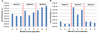
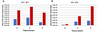
Figure 1.
Absolute peak areas of estradiol (A) and estrone (B) in three sections of a single biopsy sample, each using three chopping options before the extraction step. 1 - whole piece, 2 - chopped into large pieces, 3 - finely chopped.
|
|
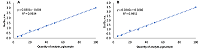
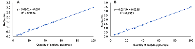
Figure 2.
Graduation curves of tested estrogens (A - estrone, B - estradiol). San - peak area of analyte, SIS - peak area of internal standard.
|

|
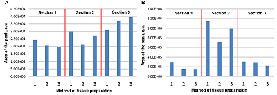
Figure 3.
The histogram of normalised peak areas of estrone and estradiol in three sections of breast tissue sample; A - normalised to tissue mass, B - normalised to lipid mass fraction. E1 - estrone, E2 – estradiol.
|

|
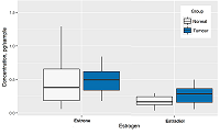
Figure 4.
The distribution of estrogen content in healthy tissue and neoplasia samples.
|
|
CLOSE

|
Table 1.
Mass spectrometric analysis conditions for each analyte.
|
|
CLOSE
|
Table 2.
Normalized estrogen peak areas for each section and chopping type. Using Student's t-test the comparison was made for each type of chopping.
|
|
CLOSE
|
Table 3.
Reproducibility and accuracy of the methodology. RSD - relative standard deviation, RME - mean error of approximation.
|
|
CLOSE
|
Table 4.
Normalised peak areas of estrone and estradiol analysed in 3 sections of a single biopsy.
|
FUNDING
This work was supported by the RFBR and the National Natural Science Foundation of China under Project No. 19-515-55021.
REFERENCES
- Holst J.P., Soldin O.P., Guo T., and Soldin S.J. (2013) Steroid hormones: relevance and measurement in the clinical laboratory, NIH Public Access, 24 (1), 105–118. DOI
- Keevil B.G. (2016) LC–MS/MS analysis of steroids in the clinical laboratory, Clinical Biochemistry, 49 (13–14), 989–997. DOI
- Russo J.R. and I.H. (2007) The role of estrogen in the initiation of breast cancer, Journal of Steroid Biochemistry and Molecular Biology, 102 (1–5), 89–96. DOI
- Hamajima N., et al. (2012) Menarche, menopause, and breast cancer risk: individual participant meta-analysis, including 118 964 women with breast cancer from 117 epidemiological studies, The Lancet Oncology, 13 (11), 1141–1151. DOI
- Dall G.V., Britt K.L. (2017) Estrogen Effects on the Mammary Gland in Early and Late Life and Breast Cancer Risk, Frontiers in Oncology, 7, 1–10. DOI
- Vermeulen A., Deslypere J.P., Paridaens R., Leclercq G., Roy F., Heuson J.C. (1986) Aromatase, 17β-hydroxysteroid dehydrogenase and intratissular sex hormone concentrations in cancerous and normal glandular breast tissue in postmenopausal women, European Journal of Cancer and Clinical Oncology, 22 (4), 515–525. DOI
- Lønning P.E., Helle H., Duong N.K., Ekse D., Aas T., Geisler J. (2009) Tissue estradiol is selectively elevated in receptor positive breast cancers while tumour estrone is reduced independent of receptor status, Journal of Steroid Biochemistry and Molecular Biology, 117 (1–3), 31–41. DOI
- Giudicessi, J. R., & Ackerman, M. J. (2013). Determinants of incomplete penetrance and variable expressivity in heritable cardiac arrhythmia syndromes. Translational Research, 161(1), 1-14. DOI
- Wudy S.A., Schuler G., Sánchez-Guijo A., Hartmann M.F. (2018) The art of measuring steroids: Principles and practice of current hormonal steroid analysis, Journal of Steroid Biochemistry and Molecular Biology, 179, 88–103. DOI
- Taylor A.E., Keevil B., Huhtaniemi I.T. (2015) Mass spectrometry and immunoassay: how to measure steroid hormones today and tomorrow, European Journal of Endocrinology, 173 (2), D1–D12. DOI
- Lee, O., Heinz, R. E., Ivancic, D., Muzzio, M., Chatterton, R. T., Zalles, C. M., ... & Khan, S. A. (2018). Breast hormone concentrations in random fine-needle aspirates of healthy women associate with cytological atypia and gene methylation. Cancer Prevention Research, 11(9), 557-568. DOI
- Laforest S., et al. (2019) Simultaneous quantification of estrogens and glucocorticoids in human adipose tissue by liquid-chromatography-tandem mass spectrometry, Journal of Steroid Biochemistry and Molecular Biology, 195 (August), 105476. DOI
- Huang H.J., Chiang P.H., Chen S.H. (2011) Quantitative analysis of estrogens and estrogen metabolites in endogenous MCF-7 breast cancer cells by liquid chromatography–tandem mass spectrometry, Journal of Chromatography B: Analytical Technologies in the Biomedical and Life Sciences, 879 (20), 1748–1756. DOI
- Blonder J., et al. (2008) Quantitation of Steroid Hormones in Thin Fresh Frozen Tissue Sections, Analytical Chemistry, 80 (22), 8845–8852. DOI
- Keski-Rahkonen P., Huhtinen K., Desai R., Tim Harwood D., Handelsman D.J., Poutanen M., Auriola S. (2013) LC-MS analysis of estradiol in human serum and endometrial tissue: Comparison of electrospray ionization, atmospheric pressure chemical ionization and atmospheric pressure photoionization, Journal of Mass Spectrometry, 48 (9), 1050–1058. DOI