The Functionalization of Calcium Phosphate Materials of Protein-Based Biologically Active Molecules
1Herzen Moscow Oncology Research Institute – branch of the National Medical Research Radiological Centre,
3 the 2-nd Botkinskiy proezd, Moscow, 125284 Russia; *e-mail: beliay@mail.ru
2Baikov Institute of Metallurgy and Material Science, Russian Academy of Science,
49 Leninskiy prospect 49, Moscow, 119334 Russia
Keywords:functionalization; biomimetic coating; biologically active molecules; octacalcium phosphate; β-tricalcium phosphate
DOI:10.18097/BMCRM00096
Recent approaches to the calcium phosphate (CaP) materials functionalization with drugs and biomolecules have been actively developed for bone defect reconstruction. However, the current techniques are low efficient in context of drug incorporation and non-controlled release from the materials. Eventually, continuous therapeutic effect in bone defect area couldn’t be achieved. The aim of this work was to develop an effective method for biologically active molecules incorporation onto the surface of CаP materials, and to study the dynamics of its release. Octacalcium phosphate (OCP), β-tricalcium phosphate (β-TCP) and β-tricalcium phosphate with biomimetic calcium phosphate layer (β-TCPmod.) were used as ceramic bioactive carriers. Bovine serum albumin (BSA) was used as a model compounds. BSA incorporation on the ceramics surface was performed by biomimetic co-precipitation from several buffer solutions containing the incorporated compound. The efficiency of biomolecules incorporation was evaluated by measuring BSA concentrations in solutions before and after materials incubation. The release of the incorporated molecules from the materials was investigated for 6 days. The structure and composition of the obtained materials were studied by application of XRD, FTIR, SEM, BET methods. It was shown that the OCP specific surface (surface area, (SBET)) was almost in 12 times higher than SBET of β-TCP. By using biomimetic approach the increase of β-TCP surface area in 1.6 times was achieved; this enhanced protein incorporation more than 3 times. The BSA biomimetic co-precipitation together with CaP on the OCP surface proved to be more effective than its adsorption from salt free solutions. The study of BSA release revealed that only 45% of loaded albumin released during 6 days of observation. Therefore, the effective method of CaP functionalization was developed. Based on biomolecules incorporation by biomimetic co-precipitation from CaP solutions, it provided a low rate of its release.

|
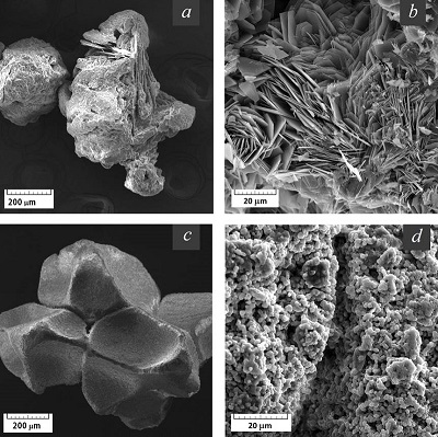
Figure 1.
SEM-photographs of the experimental materials: (a, b) – OCP, (a - х200, b - х2000),
(c, d) – β-TCP (c - х200, d – х2500). |

|
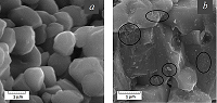
Figure 2.
Micrographs of the β-TCP ceramic surface: (a) – initial granules (х32000),
(b) – granules soaked in 2xSBF solution during 24 h (х40000). |

|
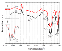
Figure 3.
FTIR spectra of the initial TCP granules (1) and soaked in 2xSBF during 24 h (2),
(a) – an overall view, (b) – detailing of the phosphate band. |
|
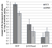
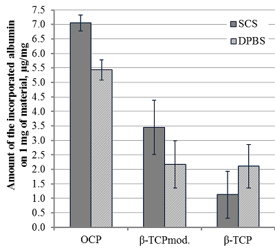
Figure 4.
Comparison of BSA loading efficacy on the OCP, β-TCPmod. and β-TCP surface with protein concentration in SCS and DPBS 3.0 mg/ml.
|
|
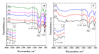
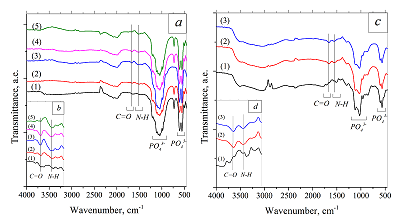
Figure 5.
FTIR spectra of materials before and after BSA functionalisation in various solutions:
(a) - β-TCP, 1 - β-TCP initial, 2 - β-TCP in SCS, 3 - β-TCPmod. in SCS, 4 - β-TCP in DPBS, 5 - β-TCPmod. in DPBS; (с) - OCP, 1- OCP initial, 2 - OCP in SCS, 3 - OCP in DPBS, (b) and (d) – detailing of FTIR-bands at 1350-1850 cm-1. |
|
CLOSE

|
Table 1.
The ionic composition of used buffer solutions.
|
|
CLOSE
|
Table 2.
Comparison of BSA amount loaded and released (for 6 days) from 1 mg of OCP.
|
|
CLOSE
|
Table 3.
Characteristics of used incorporation solutions: SCS, SBFmod., DPBS, TRIS.
|
FUNDING
The reported study was funded by RFBR within to the research project № 18-29-11052.
REFERENCES
- Bigi, A., Boanini, E. (2017) Functionalized biomimetic calcium phosphates for bone tissue repair. Journal of Applied Biomaterials and Functional Materials; 15(4), 313-325. DOI
- Esposti, L.D., Carella, F., Adamiano, A., Tampieri, A., Iafisco, M. (2018) Calcium phosphate-based nanosystems for advanced targeted nanomedicine. Journal Drug Development and Industrial Pharmacy, 44(8),1223-1238. DOI
- Parent, M., Baradari, H., Champion, E., Damia, C., Viana-Trecant, M. (2017) Design of calcium phosphate ceramics for drug delivery applications in bone diseases: A review. Journal of Controlled Release, 28(252),1-17. DOI
- Bose, S., Tarafder, S. (2012) Calcium phosphate ceramic systems in growth factor and drug delivery for bone tissue engineering: A review. Acta Biomaterialia, 8, 1401-1421. DOI
- Bigi, A., Boanini, E. (2018) Calcium phosphates as delivery systems for bisphosphonates. Journal of Functional Biomaterials, 9(1), 6. DOI
- Li, W.M., Su, C.W., Chen, Y.W., Chen, S.Y. (2014) In situ DOX-calcium phosphate mineralized Q1 CPT-amphiphilic gelatin nanoparticle for intracellular controlled sequential release of multiple drugs. Acta Biomaterialia, 15, 191-199. DOI
- Chena, G., Liuc, B., Liua, H., Zhanga, H., Yanga, K., Wanga, Q., Dingb, J., Chang, F. (2018) Calcium phosphate cement loaded with 10% vancomycin delivering highearly and late local antibiotic сoncentration in vitro. Orthopaedics and traumatology: Surgery and research, 104, 1271-1275. DOI
- Uchida, K., Sugo, K., Nakajima, T., Nakawaki, M., Takano, S., Nagura, N., Takaso, M., Urabe, K. (2018) In Vivo release of vancomycin from calcium phosphate cement. BioMed Research International, 4560647. DOI
- Peter, B., Pioletti, D.P., Lairb, S., Bujoli, B., Pilet, P., Janvier, P., Guicheux, J., Zambelli, P.-Y, Bouler, J.-M., Gauthier, O. (2005) Calcium phosphate drug delivery system: influence of local zoledronate release on bone implant osteointegration. Bone, 36, 52-60. DOI
- Barroug, A., Kuhn, L.T., Gerstenfeld, L.C., Glimcher, M.J. (2004). Interactions of cisplatin with calcium phosphate nanoparticles: in vitro controlled adsorption and release. Journal of Orthopaedic Research, 22, 703-708. DOI
- Poli, E., Magnaudeix, A., Damia, C., Lalloué, F., Chaleix, V., Champion, E., Sol, V. (2019) Advanced protocol to functionalize CaP bioceramic surface with peptide sequences and effect on murine pre-osteoblast cells proliferation. Bioorganic and Medicinal Chemistry Letters, 29, 1069-1073. DOI
- Borcard, F., Staedler, D., Comas, H., Juillerat, F.K., Sturzenegger, P.N., Heuberger, R., Gonzenbach, U.T., Juillerat-Jeanneret, L., Gerber-Lemaire, S. (2012) Chemical functionalization of bioceramics to enhance endothelial cells adhesion for tissue engineering. Journal of Medicinal Chemistry, 27, 55(18):7988-97. DOI
- Dolci L.S., Panzavolta, S., Torricelli, P., Albertini, B., Sicuro, L., Fini, M., Bigi, A., Passerini, N. (2019) Modulation of Alendronate release from a calcium phosphate bone cement: An in vitro osteoblast-osteoclast co-culture study. International Journal of Pharmaceutics, 554, 245-255. DOI
- Stigter, M., Bezemera, J., Groot, K., Layrolle, P. (2004) Incorporation of different antibiotics into carbonated hydroxyapatite coatings on titanium implants, release and antibiotic efficacy. Journal of Controlled Release, 99, 127-137. DOI
- Gurin, A.N., Komlev, V.S., Fadeeva, I.V., Barinov, S.M. (2010) Octacalcium phosphate - precursor of biomineralization, novel bone scaffold. Stomatologiia, 4, 65-72.
- Komlev, V.S., Barinov, S.M., Bozo, I.I., Deev, R.V., Eremin, I.I., Fedotov, A.Y., Gurin, A.N., Khromova, N.V., Kopnin, P.B., Kuvshinova, E.A., Mamonov, V.E., Rybko, V.A., Sergeeva, N.S., Teterina, A.Y., Zorin, V.L. (2014) Bioceramics composed of octacalcium phosphate demonstrate enhanced biological behavior. ACS Applied Materials and Interfaces, 6, 16610−16620. DOI
- Dorozhkin, S.V. (2013) Calcium orthophosphate-based bioceramics. Materials, 6 (9), 3840-3942. DOI
- Navarro, M., Michiardi, A., Castano, O., Pianeil, J.A. (2008) Biomaterials in orthopaedies. Journal of the Royal Society Interface, 5, 1137-1158. DOI
- Lin, X., Groot, K., Wang, D., Hu, D., Wismeijer, D., Liu, Y. (2015) A review paper on biomimetic calcium phosphate coatings. The Open Biomedical Engineering Journal, 9, 56-64. DOI
- Su, Y., Luo, C., Zhang, Z., Hermawan, H., Zhu, D., Huang, J., Liang, Y., Li, G., Ren, L. (2018) Bioinspired surface functionalization of metallic biomaterials. Journal of the Mechanical Behavior of Biomedical Materials, 77, 90-105. DOI
- Lee, J.Y., Choi, B., Wu, B., Lee, M. (2013) Customized biomimetic scaffolds created by indirect three-dimensional printing for tissue engineering. Biofabrication, 5, 045003. DOI
- Habibovic, P., Van Der Valk, C.M., Van Blitterswijk, C.A., De Groot, K. (2004) Influence of octacalcium phosphate coating on osteoinductive properties of biomaterials. Journal of materials science: materials in medicine, 15, 373-380.
- Shin, K., Acri, T., Geary, S., Salem, A.K. (2017) Biomimetic mineralization of biomaterials using simulated body fluids for bone tissue engineering and regenerative medicine. Tissue engineering, 23(19-20), 1169-1180. DOI
- Mirhadi, B., Mehdikhani, B., Askari, N. (2011) Synthesis of nano-sized β-tricalcium phosphate via wet precipitation. Processing and Application of Ceramics, 5(4), 193-198. DOI
- Fadeeva, I.V., Gafurov, M.R., Filippov, Ya.Yu., Davydova, G.A., Savintseva, I.V., Fomin, A.S, Petrakova, N.V., Antonova, O.S., Ahmetov, L.I., Gabbasov, B.F., Izotov, V.V., Orlinsky, S.B., Barinov, S.M. (2016) Copper-substituted tricalcium phosphates. Doklаdy Chemistry, 471(2), 384-387. DOI
- Fadeeva, I.V., Fomin, A.S., Barinov, S.M., Petrakova, N.V. (2016) Rus Patent 2578435. A61L27/02, A61L27/10, A61F2/28. The method of obtaining porous ceramics from calcium phosphates for the treatment of bone defects № 2015112518A.
- Komlev, V.S., Fedotov, A.Y. (2016) Rus Patent 2596504. A61L27/12, A61K6/00, A61F2/28, C35/447. The method of obtaining ceramics composed on octacalcium phosphate. № 2015122276.
- Termine, J.D., Posner, A.S. (1966) Infra-red determination of the percentage of crystallinity in apatitic calcium phosphates. Nature, 211, 6268.
- Tarasovich, B.N. (2012) IR spectra of the main classes of organic compounds. Reference materials. Lomonosov Moscow State University.
- Yu X., Qu, H., Knecht, D., Wei, M. (2009). Incorporation of bovine serum albumin into biomimetic coatings on titanium with high loading efficacy and its release behavior. Journal of Materials Science: Materials in Medicine, 20 (1), 287-294. DOI
- Liu, Y., Hunziker, E.B., Layrolle, P., Bruijn, J.D., De Groot, K. (2004) Bone morphogenetic protein 2 incorporated into biomimetic coatings retains its biological activity. Tissue engineering, 10(1-2), 101-108.
- Liu, Y., De Groot, K., Hunziker, E.B. (2005) BMP-2 liberated from biomimetic implant coatings induces and sustains direct ossification in an ectopic rat model. Bone, 36, 745 - 757.
- Yu, X., Qu, H., Knecht, D.A., Wei, M. (2009) Incorporation of bovine serum albumin into biomimetic coatings on titanium with high loading efficacy and its release behavior. Journal of Materials Science: Materials in Medicine, 20, 287-294. DOI
- Forsgren, J., Brohede, U., Stromme, M., Engqvist, H.K. (2011) Co-loading of bisphosphonates and antibiotics to a biomimetic hydroxyapatite coating. Biotechnology Letters, 33, 1265-1268. DOI
- Eliaz, N.,Metoki, N. (2017). Calcium phosphate bioceramics: A review of their history, structure, properties, coating technologies and biomedical applications. Materials, 10(4), 334. DOI
- Ducheyne, P., Qiu, Q. (1999) The effect of surface reactivity on bone formation and bone cell function. Biomaterials, 20, 2287-2303.
- Veresov, A.G., Putlyaev, V.I., Tretyakov, Y.D. (2004). Chemistry of inorganic biomaterials based on calcium phosphate. Russian Chemical Journal, XLVIII(4), 52-64.
- Vasserman, I.M. (1980) Chemical precipitation from solutions. L.: Chemistry.
- Barrere, F., Blitterswijk, C.A., De Groot, K., Layrolle, P. (2002) Nucleation of biomimetic Ca-P coatings on Ti6Al4V from a SBF_5 solution: influence of magnesium. Biomaterials, 23, 2211-2220.
- Barrere, F., Blitterswijk, C.A., De Groot, K., Layrolle, P. (2002) Influence of ionic strength and carbonate on the Ca-P coating formation from SBF_5 solution. Biomaterials, 23, 1921 - 1930.